iving into the pathophysiological mechanisms of myopathies associated to desmin.
Responsable du Stage : Sabrina Batonnet-Pichon
Tél : 01 57 27 79 54 E-mail: sabrina.pichon@u-paris.fr
Université Paris Cité – UMR8251 Biologie fonctionnelle et adaptative
Résumé du Projet de Stage
Desminopathies are rare neuromuscular genetic diseases with heterogeneous phenotypes and adulthood-onset. They are associated with different mutations along the gene coding for desmin. To date, the molecular mechanisms underlying desminopathies remain largely unknown and no treatment is available. Our project aims to better understand these molecular mechanisms. To this end, we have established a unique international collaboration to analyse two knock-in models carrying orthologous mutations to human R350P (R349P – R. Schroeder / C. Clemen, Germany) and R406W (R405W – S. Batonnet-Pichon, France) and the knock-out invalidated for desmin (S. Batonnet-Pichon, from Z.Li’s lab, France). These mouse models present the same phenotypic heterogeneity as in patients.
Thus, to determine the common or specific mechanisms of desminopathies, we developed a project based on 2 main axes, in which the M2 student can easily be integrated according to his/her preferences :
1/ First, to open our studies to other avenues of investigation, as the molecular mechanisms of desminopathies are poorly understood, it is essential to compare the transcriptome of our mice models to determine which new pathways could be altered in the context of myopathies. Indeed, the pathophysiology of human desminopathies is a complex, multi-scale issue with additional links to oxidative and mechanical stress response (Janué et al., 2007a, 2007b), cytoskeletal remodeling (Hakibilen et al, 2022), and protein homeostasis (Winter et al., 2018). Notably, a recently reported transcriptomic analysis performed on muscle tissue derived from horses affected by myofibrillar myopathies showed alterations in cysteine metabolic pathways (Valberg et al., 2018). Since we have the exclusive opportunity to compare three desminopathy mouse lines, i.e. our R405W and R349P KI models and the KO mouse, we have already perform RNAseq, with ICM platform, from soleus muscles of WT, heterozygous or homozygous mice of each model. Analysis and comparison are in progress. Moreover proteomics analysis will be performed in the next months. We expected to discover new deregulated pathways, and therefore to confirm these results with different approaches as qRT-PCR, IHC, WB on mice models or corresponding CrisPR modified human myoblasts.
2/ In parallel, we aim to investigate if the regeneration process is affected in our mice models. It has already been described that the lack of desmin leads to an altered regeneration process with delay in fiber repair and fibrosis formation. However, this aspect has never been addressed in the context of desminopathies due to missense mutations. Notable, our preliminary data showed that homozygous R405W KI mice have a large percentage of central myonuclei at the age of 3 months (around 35 % compared to 2 % in wt). In the same way heterozygous animals also present a moderate increase (around 20 %) of central myonuclei from 3 to 12 months of age. This finding is indicative of an active regeneration process starting already at an early age. Corresponding analysis will now be performed in R349P mice.
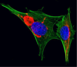
Satellite cell regeneration and differentiation are generally closely linked to adhesion and migration processes. Here it is noteworthy, that we have already demonstrated that satellite cells extracted from homozygous R405W KI as well as KO mice displayed a decrease in cell adherence under shear stress and an increase of migration velocity on fibronectin and laminin (Hakibilen et al, 2022) coated surfaces.
Moreover, we have recently obtained human immortalized myoblast modified by CRISPR/Cas9 either invalidated for desmin, or with specific mutation (cell screen in progress). Since the actin cytoskeleton is crucial for cell adhesion and migration, we will determine the organization of actin stress fibers in satellite cells extracted from all our mouse models and/or human myoblasts using a micropatterning approach (in collaboration with S. Hénon, Paris University). This will enable us to analyse others adhesion and contractility markers like the direct desmin interaction partner synemin. Furthermore, we will generate 3D muscle-microtissues in collaboration with S. Hénon (MSC, Paris university) and T. Boudou (LiPhy, Grenoble). This system will allow us to follow the kinetic of formation of the microstissue via videomicroscopy. In collaboration with M.Reffay, we have also the possibility to form 3D magnetic spheroids (Nagle et al, submitted). Therefore we will check markers of adhesion as cadherin for cell-cell interaction, or synemin in context of desmin mutant expression compared to WT cells. We also can measure the strength develop by the microtissue with the cell capacity to deform the elastic plot. In addition, we will also be able to follow all these markers during cell differentiation.
Finally, to determine the effect of mutant desmin on the adhesion and migration in vivo, we want to adapt a unique technique that permits to follow satellite cell migration within living muscle tissue (Ishido and Kasuga, 2011). By following the migration of satellite cells that are either prepared from wild-type mice or our desminopathy mouse models and subsequently transfected with a luciferase marker, we will be able to clarify the question if satellite cell migration is affected in the context of desminopathy-causing mutations.
In conclusion, this work is important to better understand the differential mechanisms that rule the fate of desmin mutant in patients, and help to define strategies for future treatments. Indeed, using the crosstalk between our previous cellular and the planed comparison of our mouse models, we could predict what type of drug to screen for future treatments. Overall, this project will establish a bridge between patients and molecular mechanisms involved in desminopathies.
Dernières Publications en lien avec le projet :
- Hakibilen C., Delort F., Daher M.T., Joanne P., Cabet E., Cardoso O., Bourgois-Rocha F., Tian C., Rivas E., Madruga M., Ferreiro A., Lilienbaum A., Vicart P., Agbulut O., Hénon S.and Batonnet-Pichon S. Desmin modulates muscle cell adhesion and migration. Cell Dev. Biol., March 8. 2022. doi: 10.3389/fcell.2022.783724
- Delort F, Segard BD, Hakibilen C, Bourgois-Rocha F, Cabet E, Vicart P, Huang ME, Clary G, Lilienbaum A, Agbulut O, Batonnet-Pichon S. Alterations of redox dynamics and desmin post-translational modifications in skeletal muscle models of desminopathies. Exp Cell Res. 2019 Oct 15;383(2):111539. doi: 10.1016/j.yexcr.2019.111539.
- Charrier EE, Montel L, Asnacios A, Delort F, Vicart P, Gallet F, Batonnet-Pichon S, Hénon S. The desmin network is a determinant of the cytoplasmic stiffness of myoblasts. Biol Cell. 2018 Apr;110(4):77-90. doi: 10.1111/boc.201700040.
- Even C, Abramovici G, Delort F, Rigato AF, Bailleux V, de Sousa Moreira A, Vicart P, Rico F, Batonnet-Pichon S, Briki F. Mutation in the Core Structure of Desmin Intermediate Filaments Affects Myoblast Elasticity. Biophys J. 2017 Aug 8;113(3):627-636. doi: 10.1016/j.bpj.2017.06.020.
- Barateau A, Vadrot N, Agbulut O, Vicart P, Batonnet-Pichon S, Buendia B. Distinct Fiber Type Signature in Mouse Muscles Expressing a Mutant Lamin A Responsible for Congenital Muscular Dystrophy in a Patient. 2017 Apr 24;6(2):10. doi: 10.3390/cells6020010.
Ce projet s’inscrit-il dans la perspective d’une thèse :
oui x
si oui type de financement prévu : Ministère
Ecole Doctorale de rattachement : BioSPc
Intitulé de l’équipe : Myologie fondamentale et tranlationnelle
Intitulé de l’Unité : UMR 8251 Biologie Fonctionnelle et adaptative
Nom du Responsable de l’Unité : Jean-Marie Dupret
Nom du Responsable de l’Équipe : Ana Ferreiro
Adresse : Université Paris Cité BFA, bâtiment Lamarck 35 rue Hélène Btion 75205 Paris cedex 13